Acne Detection Methods and Performance Analysis using Component Images of Various Color Spaces
Copyright ⓒ 2021 The Digital Contents Society
This is an Open Access article distributed under the terms of the Creative Commons Attribution Non-CommercialLicense(http://creativecommons.org/licenses/by-nc/3.0/) which permits unrestricted non-commercial use, distribution, and reproduction in any medium, provided the original work is properly cited.

Abstract
In previous studies for acne detection, methods using component images related to redness in the RGB, HSV, and CIE L*a*b* color spaces have been proposed. Component image R in RGB color space and gray-level image, H and V in HSV, and a* in CIE L*a*b* were applied. In the proposed acne detection, component images R and Cr in RGB and YCbCr color space were used. The component image Cr is a difference from the reference value of the red components, and is suitable for the redness information analysis for acne detection. In this paper, we analyzed the performance of previous studies and the proposed acne detection method, and the proposed method detects acne more effectively than the existing methods. This image processing-based acne detection should not be the purpose of diagnosis, but will help reduce the excessive effort of dermatologists who manually check acne.
초록
여드름 검출을 위한 선행연구들에서는 RGB, HSV, CIE L*a*b* 컬러 공간에서 적색과 관련된 성분 영상들을 이용한 방법들이 제안되었다. RGB 컬러 공간에서는 R과 변환된 그레이레벨, HSV에서는 H와 V, CIE L*a*b*에서는 a*의 성분 영상들이 적용 되었으며, 제안하는 여드름 검출은 RGB와 YCbCr 컬러 공간에서의 성분 영상 R과 Cr을 이용하였다. 성분 영상 Cr은 적색 성분들의 기준값과의 차이를 나타내므로 여드름 검출을 위한 적색의 화소 정보 분석에 적합하다. 본 논문에서는 선행연구들과 제안하는 방법의 여드름 검출의 성능분석을 수행하였으며, 제안하는 방법이 기존의 방법들보다 효과적으로 여드름을 검출하였다. 이러한 영상처리 기반의 여드름 검출은 진단 목적이 아닌 여드름을 수동으로 확인하는 피부과 의사들의 과도한 노력을 감소시키는데 도움이 될 것이다.
Keywords:
Acne Detection, Skin Disease Analysis, Beauty Clinic, Skin Care, YCbCr Color Model키워드:
여드름 검출, 피부질환 분석, 뷰티 클리닉, 피부 관리, YCbCr 칼라 모델Ⅰ. Introduction
Today, various skin care for clean skin is recognized as part of physical or mental health care [1-3]. For this purpose, numerous beauty clinic programs are being used to treat skin diseases such as skin tone(or color), pores, acne, and pigmentation, regardless of age or gender. In particular, in the previous study [4], a survey was conducted on 302 female college students to analyze their concerns about skin care. As a result of the survey, skin diseases such as pores, skin tone, acne and excessive sebum (or oils) were analyzed as 32.1%, 22.5%, 18.5%, and 9.9%, respectively.
Skin tone and pigmentation, a phenomenon that occurs when the amount of pigment in the skin tissue changes abnormally, is related to melanin and hemoglobin pigments. In general, skin tone is determined by the amount of melanin, and as the amount of melanin increases, the skin tone becomes darker. The detection of images corresponding to melanin from digital images was proposed in a previous study [5]. In order to analyze pores, a digital image is generally obtained using a photographing device equipped with a microscope and a telemedicine haptic. However, in the previous study [6], a method of photographing and analyzing pores based on an image sensor built into a smartphone without a separate microscope was also proposed.
In this paper, a method for acne detection in skin diseases was proposed, and a comparative analysis was performed between the previous studies and the proposed method. In fact, skin disease diagnosis should be made by a dermatologist, but the previous studies and suggested acne detection will serve as an effective assistant to reduce time in the process of manually checking and diagnosing skin diseases.
The rest of the paper is organized as follows. In Section 2, the acne detection methods of previous studies are presented, and the proposed acne detection is explained in Section 3. In Section 4, the performance of previous studies and the proposed method was compared and analyzed. Finally, Section 5 describes the conclusions and future studies.
Ⅱ. Previous Studies for Acne Detection
In this section, color models and algorithms adopted in previous studies for detecting acne candidate regions in images containing acne are described. However, in the acne detection method of previous studies, a process such as detecting a face region or a region of interest (ROI) is not described.
2-1 Acne detection using R, G, B channels
In the previous study [7], automated lesion counting for 5 types of acne such as nodule, pustule, papule, whitehead and blackhead comedone was performed based on image processing. Here, the authors evaluate that the sensitivity and predictive value of the lesion counting program has more than 70% performance compared to the proposed method and manual counting performed by a professional dermatologist [7]. The color space applied for acne detection is based on the basic RGB color space, and the detection process is very simple as shown in Equations (1) and (2).
| (1) |
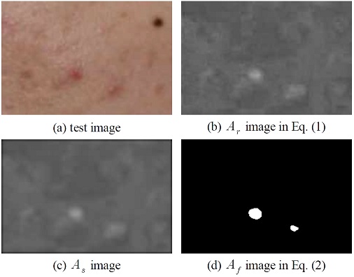
In Equation (1), Ar is an equation for calculating a region (or channel) with a large influence of red, and a smooth image As is obtained from the obtained Ar through filtering with a disk average. Then, as shown in Equation (2), the final acne candidate As was detected by comparing the obtained image Af with the threshold value T, and (i, j) represents the pixel position of the image.
| (2) |
Figure 1 shows the results of acne detection obtained by Equations (1) and (2) and the main result images for each stage. In Figure 1, the threshold T for acne detection was set at a pixel value of 120. As described so far, the method of the previous study [7] is very simple and has the advantage of fast calculation.
2-2 Acne detection using Gray-level and V channel
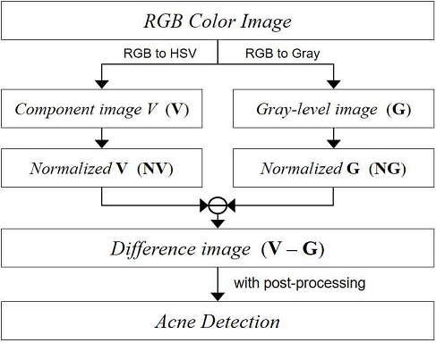
Acne detection in the previous study [8] was used based on the gray-level space of the RGB color image and the component image V of the converted HSV color space, and the overall flow of acne detection is shown in Figure 2. First, a gray-level image and a component image V are obtained, and then normalization is performed on the two images with values between 0 and 1 as in Equation (3).
| (3) |
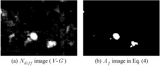
where img includes the two images V and G in Figure 2, and the final Nimg represents the two normalized images NV and NG. Then, the difference image Ndiff between the two images was acquired and adjusted, and the finally detected acne candidate Af was detected by Equation (4). The authors set the threshold value T in Equation 4 to 0.75, which is a double type value.
| (4) |
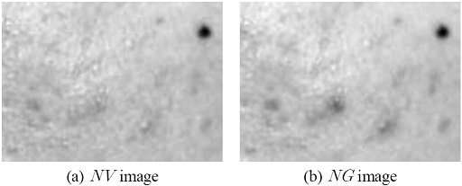
Figure 3 shows the normalized images NV and NG from the image of Figure 1(a), and Figure 4 shows the final acne candidate image detected by the method of the previous study [8].
2-3 Acne detection using RGB and HSV color model
In the previous study [9], each component image of the RGB color space and H (hue) image of the HSV color space were used for acne detection.
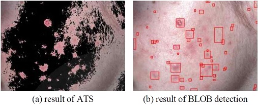
First, the smooth Gaussian method was pre-processed on the component image G of the RGB color space, and the image was segmented using the adaptive thresholding segmentation (ATS). And then, acne was quantified using a simple BLOB detector and speed up robust feature (SURF) technology. In addition, the redness included in the image applies the range of 0 to 8 and 172 to 255, which is the red color range of H. As the main features for acne quantification, the standard deviation (SD) of each component image in RGB color space, redness, mean of H, mean and SD of S (saturation), and Shannon entropy were used. On the basis that acne is generally circular, the circularity [9, 10] as in Equation (5) was also applied as a feature for quantification.
| (5) |
where A and P are the area and perimeter of the blob, respectively. Figure 5 shows the result images of reference [9].
2-4 Acne detection using RGB and CIE color model
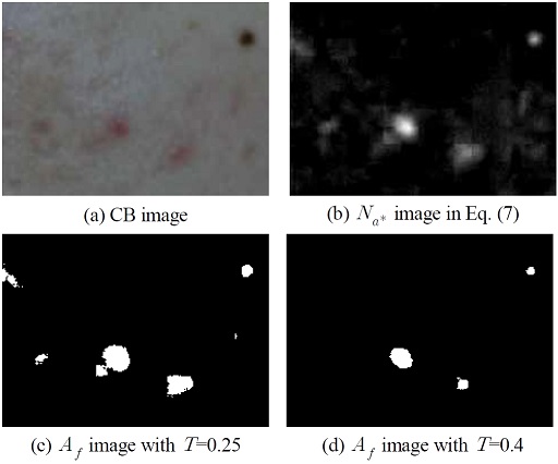
Acne detection in the previous study [11] used the component image a* after converting the RGB color space into the CIE L*a*b* color space. First, a color balancing (CB) of test image was performed using Equation (6) as the pre-processing process, and a color space was converted from RGB to CIE L*a*b*.
| (6) |
where Ni represents a value inverted by the average of each component image, and Si is a scaling factor of each component image normalized to the maximum value of Ni = R, G, B. Therefore, each component image of the color balanced image is acquired as R×SR, G×SG, and B×SB. After converting the color balanced image into CIE L*a*b*, acne was detected using the component image a*.
The authors used the feature that when the value of component image a* is negative, it is green, and when the value is positive, it is close to red. Based on these features, the acne detection was performed by Equation (7). Figure 6 shows the acne detection results of reference [11] using the test image of Figure 1(a).
| (7) |
Ⅲ. Proposed Method for Acne Detection
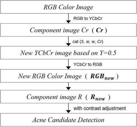
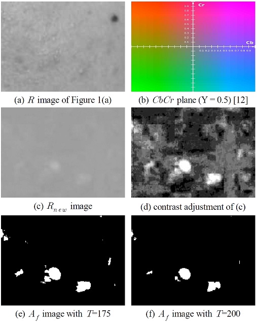
The overall flow of the proposed acne detection is shown in Figure 7, and the component image Cr is used after converting the RGB color image into the YCbCr color space. Since acne is generally red, the acne region can be inferred using pixel information of the component image R. However, as shown in Figure 8(a), acne cannot be estimated using only the information of component image R, that is, brightness information. In other words, all regions such as acne, spots, and pigmentation can be detected. Therefore, the component image Cr was used to separate the red information from the information of the component image R. Since the component image Cr shows the difference from the reference value of the red components, it is suitable for acne detection.
First, if the constant luminance (Y) value is set to 0.5 in the YCbCr color space, the CbCr plane is shown in Figure 8(b) [12]. Based on this, a new YCbCr color space is created as shown in Equation (8).
| (8) |
where the parameter w is set to the median of the pixel range, and is 128. Then, the obtained YCbCrnew is converted back to the RGB color space to generate a new image RGBnew. The component image Rnew extracted from the generated RGBnew is shown in Figure 8(c). Figure 8(d) is the result of applying the contrast adjustment between 0 and 1 to the image of Figure 8(c). The acne detection is detected by Equation (9), and when the threshold value T is 200 or more, it is determined as a red region.
| (9) |
Figures 8(e) and (f) show the results of setting the threshold value T to 175 and 200 in Equation (9), respectively. In this paper, we estimated that if the pixel values in Figure 8(d) were between 175 and 200, it was pigmentation, and if it was 200 or more, it was acne. This is because even the same red color can be separated into pigmentation and acne.
Ⅳ. Experiment and Discussion
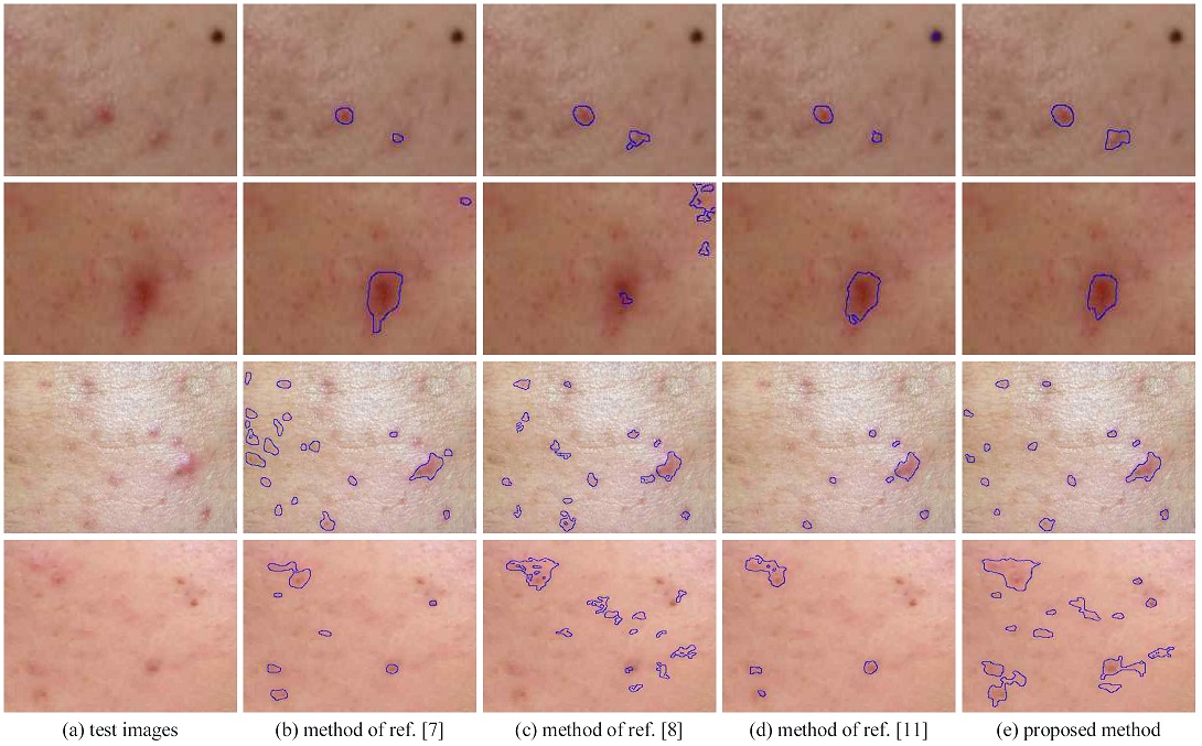
In this section, the performance of the previous studies [7, 8, 11] described in Section 2 and the proposed method were compared and analyzed. In the pre-processing and acne detection process, acne candidate regions were detected by Equation (2, 4, 7), which is each method of previous studies, and Equation (9) which is the proposed method. Figure 9 shows the results of the final acne detection in each study. However, the post-processing procedure after detecting acne candidate regions is different among previous studies.
Therefore, the post-processing procedure was unified for objective performance analysis. In the post-processing, objects smaller than 50 in size were removed from the binary image including the acne candidate region, and the morphological imaging techniques of erosion and dilation operations were applied for noise removal. At this time, a mask for the operation was applied in the form of a disk with the size of 3 by 3.
In Figure 9, the threshold value T applied to reference [7] was set to 120, 145, 100, and 147, from top to bottom, respectively. This means that the threshold value T must be applied differently according to the brightness of the input image. In the acne detection result of reference [8], the second image was detected incorrectly because the image was dark and ruddy as a whole, and the brightness information of the selected gray-level and component image V was similar. The result of reference [11] is generally good, but a spot was also detected in the acne detection result of the first image. Table 1 shows the color models and component images adopted in the methods which is described in this paper for the acne detection. Only RGB color model was used in reference [7], and component images R and Cr were applied in the proposed method.
The detection of various skin diseases, including acne, should not be used for diagnostic purposes. Image processing-based acne detection should be a means to help overcome the excessive effort of dermatologists who manually check for acne.
Ⅴ. Conclusions and Future Works
In this paper, the general characteristic that acne is red (or ruddy) is applied to the proposed acne detection as in previous studies. The main procedure is to obtain YCbCr color space where the intensity of constant luminance and Cb are 0.5 after converting RGB color space-based image into YCbCr color space. Then, it is converted back to the RGB color space. The acne regions were detected using the component image R in the converted RGB color space. In this image, if it is greater than the threshold value of 200, it was estimated as acne. As a result of the experiment, it was confirmed that the proposed method is more effective in detecting acne than the existing methods. However, the acne detection using image processing should never make a final diagnosis, and should be a means to help overcome the excessive efforts of dermatologists. In the future, we plan to conduct deep learning-based research for skin analysis such as the frontal face detection, pigmentation, blemishes, and wrinkles.
Acknowledgments
This work was supported by Hanshin University Research Grant.
References
- Y. B. Kim, J. H. Baek and B. C. Ahn, “An Implementation of a Skin Care System for Android Phones,” in Proceedings of International Conference on Embedded Systems, Cyber-physical Systems, & Applications, pp. 57-61, 2016.
- J. D. Kim and J. E. Seol, “A Study on the State of Acne Awareness and Care,” The Journal of the Korean Society of Knit Design, Vol. 13, No. 2, pp. 1-9, June, 2015.
-
K. H. Park and Y. H. Kim, “Skin Condition Analysis of Facial Image using Smart Device - Based on Acne, Pigmentation, Flush and Blemish,” Journal of Advanced Information Technology and Convergence, Vol. 8, No. 2, pp. 47-58, Dec. 2018.
[https://doi.org/10.14801/JAITC.2018.8.2.47]

- J. D. Kim, “Study on Female College Students’ Recognition and State on Skin care,” The Journal of Korean Society of Knit Design, Vol. 15, No. 1, pp. 54-65, Feb. 2017.
-
L. Yang, S. H. Lee, S. G. Kwon and K. R. Kwon, “Skin Pigmentation Detection Using Projection Transformed Block Coefficient,” Journal of Korea Multimedia Society, Vol. 16, No. 9, pp. 1044-1056, Sep. 2013.
[https://doi.org/10.9717/kmms.2013.16.9.1044]

- J. S. Bae, J. H. Jeon, J. Y. Lee and J. O. Kim, “Skin Condition Estimation Using Mobile Handheld Camera,” ETRI Journal, Vol. 38, No. 4, pp. 776–786, August, 2016.
-
S. Min, H. J. Kong, C. Yoon, H. C. Kim and D. H. Suh, “Development and evaluation of an automatic acne lesion detection program using digital image processing,” Skin Research and Technology, Vol. 19, pp. e423-e432, 2013.
[https://doi.org/10.1111/j.1600-0846.2012.00660.x]

-
T. Chantharaphaichit, B. Uyyanonvara, C. Sinthanayothin, and A. Nishihara, “Automatic Acne Detection for Medical Treatment,” in Proceeding of the 6th International Conference of Information and Communication Technology for Embedded Systems (IC-ICTES), Hua Hin, Thailand, pp. 33-38, 2015.
[https://doi.org/10.1109/ICTEmSys.2015.7110813]

- N. Kittigul, Automatic Acne Detection and Quantification for Medical Treatment through Image Processing, M.S. dissertation, Sirindhorn International Institute of Technology, Thammasat University, Bangkok, 2017.
- LearnOpenCV, Blob Detection Using OpenCV [Internet]. Available: https://learnopencv.com/blob-detection-using-opencv-python-c/, .
-
K. H. Park and H. S. Noh, “Effective Acne Detection using Component Image a* of CIE L*a*b* Color Space,” Journal of Digital Contents Society, Vol. 19, No. 7, pp. 1397-1403, July 2018.
[https://doi.org/10.9728/dcs.2018.19.7.1397]

- Wikipedia, YCbCr [Internet]. Available: https://en.wikipedia.org/wiki/YCbCr, .

2007: Dept. of IT Engineering, Graduate School of Mokwon University (Ph.D Degree)
2012: Graduated School of Engineering, Fukuoka Institute of Technology (Ph.D Degree)
2007~2009: Senior Engineer, Technology Lab., Fumate Co., Ltd.
2009~2011: Research Professor, 2nd BK21, Chosun University
2012~2021.02: Assistant Professor, Div. of Convergence Computer & Media, Mokwon University
2021.03~now: Assistant Professor, Div. of Computer Engineering, Hanshin University
※Research Interests : Disaster prevention application and technology, ICT Convergence, Vehicle-IT, IoT, UWB, Digital wireless communication, etc.

2005: Dept. of IT Engineering, Graduate School of Mokwon University (M.S Degree)
2010: Dept. of IT Engineering, Graduate School of Mokwon University (Ph.D Degree)
2008~2009: Researcher in Prevention of Disaster with Information Technology RIC.
2011~2012: Senior Researcher in Inconex Co., Ltd.
2012~now: Assistant Professor in Div. of Convergence Computer & Media, Mokwon University.
※Research Interests : digital content, computer vision, pattern recognition, H.26x, aviation application technology, disaster prevention application and technology, etc.
